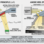
Off-focus radiation (also known as extrafocal radiation) refers to X-ray photons produced outside the designated focal spot on the target track of an X-ray tube assembly. While ideal X-ray tube design dictates that high-velocity electrons interact exclusively within the boundaries of the focal spot (determined by the cathode focusing cup), a fraction of incoming electrons bounce off the target surface and strike other parts of the anode assembly.
This secondary interaction produces an unwanted, diffuse X-ray flux that degrades image quality, reduces contrast, increases patient radiation exposure, and introduces characteristic imaging artifacts.
1. Physical Mechanism & Origin
When high-velocity incident electrons strike the tungsten target at the focal spot, not all of their kinetic energy is immediately converted into X-rays or heat through Bremsstrahlung and Characteristic interactions.
- Electron Scattering: A percentage of primary electrons undergo high-angle elastic collisions and bounce (rebound) off the focal spot area back into the vacuum space inside the glass or metal envelope.
- Re-Acceleration: Because the anode assembly maintains a strong positive electric potential relative to the cathode, these free-floating secondary electrons are pulled back toward the anode disc, stem, or target area outside the actual focal spot.
- Extrafocal Emission: Upon impacting these non-target tungsten or molybdenum structures, the secondary electrons undergo Bremsstrahlung interactions, generating off-focus X-ray photons.
(Primary Rays)
2. Technical and Clinical Impacts
Off-focus radiation accounts for 10% to 15% of the total primary beam flux in unshielded diagnostic X-ray tubes. Its presence creates three major diagnostic and radiation safety problems:
A. Loss of Image Contrast and Spatial Resolution
- Off-focus photons exit the X-ray tube at wide, unpredictable angles relative to the central ray.
- These extrafocal photons bypass the primary collimator leaves and expose areas of the patient and image receptor outside the intended field of view.
- This adds a background “fog” or noise layer to the image, significantly lowering overall image contrast and obscuring fine anatomical details (loss of spatial resolution).
B. Geometric Artifacts (“Ghosting”)
- Off-focus radiation can project anatomical structures located outside the collimated area onto the final radiograph.
- This creates a characteristic shadow or “ghosting” artifact around soft tissue borders and high-density bone margins (e.g., bone edges appearing doubled or blurred beyond collimation lines).
C. Increased Patient Radiation Dose
- Extrafocal photons deliver unnecessary skin and tissue dose to areas outside the primary region of interest (ROI).
Image Quality
Decreased Contrast & Sharpness
Artifacts
Extrafocal “Ghosting” Beyond Collimation
Radiation Safety
Unintended Patient Exposure Outside ROI
3. Engineering Solutions and Control Measures
Modern X-ray tube housing and collimator assemblies incorporate structural design elements to absorb and eliminate off-focus radiation before it reaches the patient.
(Positioned close to Output Window)
Engineering Interventions:
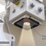
- Fixed Lead Diaphragm / Aperture (First Stage Collimation): Positioned immediately below the X-ray tube output window inside the housing, a fixed lead diaphragm absorbs wide-angle off-focus rays near their origin.
- Metal-Envelope Tubes (Grounded Metal Housing): Modern high-output diagnostic tubes replace traditional glass envelopes with metal-backed structures maintained at a constant electrical potential. The metal wall conducts scattered secondary electrons away, preventing them from returning to strike the anode.
- Dual-Stage Adjustable Collimator Blades: Primary collimator shutters placed closest to the tube port absorb extrafocal rays, while secondary lower shutters shape the final primary beam field.
4. Summary of Key Differences
| Property | Primary Focal Radiation | Off-Focus Radiation |
|---|---|---|
| Point of Origin | Actual Focal Spot on Anode Target Track | Anode Disc Surface, Target Stem, or Rotor Structure |
| Electron Source | Direct Cathode Filament Electrons | Rebounded / Backscattered Electrons |
| Beam Alignment | Collimated, Predictable Geometry | Wide-Angle, Divergent Scatter |
| Clinical Effect | Forms Diagnostic Image Signal | Creates Fog, Reduces Contrast, Increases Patient Dose |
| Control Method | Line-Focus Principle & Collimation | Internal Lead Diaphragms & Metal-Envelope Tubes |
Summary
Off-focus radiation is an unwanted byproduct of electron scattering within the X-ray vacuum tube. Left unmanaged, extrafocal photons degrade image quality and deliver non-diagnostic radiation dose to the patient. By pairing metal-envelope tube design with internal fixed lead diaphragms and stage-one collimation, radiographers and engineers effectively suppress off-focus radiation to maintain high-contrast diagnostic images.