What is Radiation Detection? Principles, Efficiency, and Radiation Detector Types

Radiation detection and measurement form the absolute backbone of radiology, radiation therapy, nuclear medicine, health physics, and industrial safety compliance. Because ionizing radiation is entirely imperceptible to normal human senses—such as sight, hearing, touch, or smell—specialized scientific instruments and precise detection methodologies are mandatory to locate, quantify, and regulate exposure levels safely.

1. Fundamental Physical Principles of Radiation Detection
Radiation interacts with matter by depositing energy at a microscopic subatomic level, triggering several distinct physical, chemical, and biological phenomena that serve as the fundamental basis for all detection instruments.
Ionization Phenomenon: High-energy ionizing radiation (such as alpha particles, beta particles, gamma rays, and X-rays) transfers enough energy to neutral atoms or molecules to strip away orbital electrons. This creates positive ions and free electrons, known collectively as ion pairs. By establishing an external electric field inside a chamber, these liberated charges are swept toward electrodes, producing measurable electrical pulses or continuous currents
Luminescence and Scintillation: When radiation strikes certain crystalline or organic materials (phosphors), their constituent atoms are elevated to excited energy states. As these atoms return to their ground state, they release the stored energy in the form of flashes of visible or ultraviolet light. When paired with light-sensitive photomultiplier tubes (PMTs), these light photons are converted into amplified electrical signals.
Thermoluminescence (TLD Process): Certain high-purity crystalline solids (such as lithium fluoride, , or calcium sulfate, ) and specialized glasses possess crystal lattice defects. When exposed to radiation, electrons become trapped within these forbidden energy states. When the material is subsequently heated in a controlled laboratory reader, the trapped electrons return to the ground state, releasing the stored energy as luminescent light proportional to the absorbed radiation dose.
Photographic and Chemical Alterations: Similar to visible light, ionizing radiation interacts with silver halide crystals in photographic emulsions to create a latent image composed of stable black metallic silver grains upon chemical development. Furthermore, radiation-induced chemical shifts—such as the oxidation of ferrous sulfate to ferric sulfate in Fricke dosimeters or distinct color changes in specialized radiochromic plastics—allow for high-dose tracking in industrial irradiators.
Biological Biodosimetry: In catastrophic scenarios where physical dosimeters are absent or compromised, biological assessment techniques serve as a vital fallback. Analyzing peripheral blood lymphocytes for structural chromosomal aberrations (such as dicentric chromosomes or rings) provides a reliable retrospective evaluation of radiation exposure across high dose ranges ().
2. Advanced Signal Processing Dynamics: Pulse Mode vs. Current Mode
Because the quantum packets of energy deposited by individual radiation interactions are exceptionally minuscule, detectors require sophisticated electronic circuits for pulse shaping, amplification, and processing. Depending on the intensity of the radiation field and the application requirements, systems operate under two distinct operational paradigms:
Pulse Mode Operation: In pulse mode, the detector processes each individual particle or photon interaction as an isolated, discrete event. Every physical strike generates an independent electrical pulse whose amplitude is typically proportional to the energy deposited by that specific interaction.
The Dead Time Phenomenon: A critical operational parameter in pulse mode is dead time—the minimum finite time interval required between two consecutive ionizing interactions for the electronic system to register them as two separate, distinct signals.
Paralyzable vs. Non-Paralyzable Systems: In a non-paralyzable detector, any event arriving during the dead time is simply ignored without extending the lockout window. In a paralyzable detector, each incoming event (even those missed during dead time) extends the recovery window, causing extreme high-flux radiation fields to paradoxically drive the registered count rate down toward zero. Consequently, pulse mode is ideal for low-to-moderate radiation environments (such as contamination surveys and spectroscopy).
Current Mode Operation: When radiation interaction rates and particle flux become excessively high (such as inside active radiotherapy treatment rooms or near nuclear reactor cores), pulse mode fails because individual pulses overlap continuously, resulting in severe dead time losses and unreadable output.
Integration Mechanics: To overcome this, current mode operation continuously integrates and averages the electrical signals generated by millions of overlapping individual interactions.
Output Metric: Instead of counting individual pulses, the system measures a steady net direct current (DC) output. This net current is directly proportional to the aggregate dose rate or energy fluence deposited per unit time within the detector volume, ensuring stable and reliable measurement without saturation bottlenecks in intense radiation fields.
3. Evaluating Detector Performance and Efficiency Metrics
The overall efficacy and performance capability of any radiation detector are quantified through specific efficiency metrics that determine how successfully incoming radiation is captured and recorded.
Geometric Efficiency (): This metric represents the spatial probability fraction of radiation emitted by a radioactive source that actually intercepts the active window or sensitive volume of the detector. It depends heavily on the physical distance between source and detector, as well as their relative orientation:
Intrinsic Efficiency / Quantum Detection Efficiency (): This measures the internal conversion probability—the fraction of radiation particles or photons that actually interact with the detector material once they have successfully reached its sensitive volume:
Intrinsic efficiency is heavily influenced by the physical thickness of the detector, material density, effective atomic number (), and the incident photon energy. High-density, high- materials absorb penetrating gamma rays much more efficiently.
Total Detector Efficiency (): The overall performance product combining spatial geometry and internal conversion capability:
4. Major Classification of Radiation Detectors
Radiation detection systems are systematically categorized based on their working medium and primary detection mechanism into three major classes:
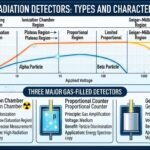
Gas-Filled Detectors: Utilize a gas volume enclosed between electrodes. By adjusting the applied voltage, these operate across three distinct operational regions:
Ionization Chambers: Operated at low voltage to collect primary ion pairs without gas multiplication; ideal for high-intensity dose-rate measurements.
Proportional Counters: Operated at moderate-to-high voltage to induce gas amplification (avalanche effect), enabling precise particle discrimination (alpha vs. beta).
Geiger-Muller (GM) Counters: Operated at high voltage where a single ionizing event triggers a full-volume gas discharge avalanche, providing extreme sensitivity for portable survey meters.
Scintillation Detectors: Combine a transparent luminescent crystal (such as Thallium-activated Sodium Iodide, ) with a Photomultiplier Tube (PMT) or photodiode. They offer high intrinsic efficiency for gamma rays and are widely deployed in nuclear medicine imaging cameras and spectroscopic analysis.
Semiconductor (Solid-State) Detectors: Utilize high-purity solid semiconductor crystals (such as High-Purity Germanium, , or Silicon) instead of gas or liquid media. Radiation creates mobile electron-hole pairs. Because the energy required to create an electron-hole pair is extremely low compared to gas ionization, semiconductor detectors deliver unmatched energy resolution for precision gamma spectrometry.
5. Mathematical Framework Summary
Radioactive Decay Law:
(Where is initial activity, is the decay constant, and is elapsed time)
Dose Equivalent Equation:
(Where is absorbed dose in Grays and is the radiation weighting factor)
6. Comparative Analysis Table