Ionization Chambers: Engineering, Dosimetry, QA/QC, and Clinical Applications

Ionization chambers represent the gold standard in radiation dosimetry, offering exceptional long-term stability, high precision, and reliability. They are deployed extensively across diagnostic radiology, radiation therapy physics, health physics, and industrial radiation safety compliance. Operating on the principle of direct gas ionization without internal multiplication, ionization chambers provide robust quantitative measurement across a wide spectrum of ionizing radiation fields.
1. Fundamental Working Principles and Ionization Mechanics
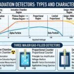
An ionization chamber operates within the saturation plateau region (Region II) of the gas-filled detector voltage response curve, ensuring complete collection of all liberated charges.
- Ion Pair Generation and Energy Dynamics: When ionizing radiation traverses the active gas volume, it transfers kinetic energy to gas molecules via inelastic collisions. For an air-filled chamber, an average of 34 eV of energy is required to produce a single ion pair. For instance, a single 100 keV photon interacting within the active volume can create approximately 3000 ion pairs.
- Voltage Independence and Signal Amplitude: Once radiation-induced ion pairs (positive ions and free electrons) are formed, an external electric field sweeps them toward their respective electrodes. The amplitude of the resulting signal depends entirely upon the total number of ion pairs formed and remains strictly independent of the applied voltage across the saturation plateau.
- Bragg-Gray Cavity Principles: If a chamber is filled with air and the effective atomic number (Z) of its outer wall material matches that of air, the ionization current produced becomes directly proportional to the exposure rate, measuring the average rate of energy dissipation within the cavity volume.
2. Structural Design and Construction Components
A standard ionization chamber is engineered with specific structural elements designed to ensure measurement precision:

- The Outer Shell (Cathode): Typically constructed as a cylindrical enclosure whose inner wall is coated with a thin layer of conducting material—such as graphite—to make it electrically conductive and ensure uniform electric field distribution.
- The Central Electrode (Anode): A rigid metal rod or high-purity wire acting as the collecting electrode, positioned along the central axis and meticulously insulated from the grounded outer chamber wall.
- Fill Gas and Density Dynamics: The internal volume can be sealed or vented.
- Air-Vented Chambers: Communicate freely with ambient barometric pressure, requiring strict temperature-pressure correction factors (Pth).
- High-Z / Pressurized Fillings: To overcome the inherently low intrinsic efficiency caused by low gas density, chambers are frequently filled with high atomic number noble gases like argon (Z=18) or xenon (Z=54), or pressurized to significantly increase sensitivity toward diagnostic X-rays and gamma rays.
3. Major Operational Modes
Ionization chambers operate under two primary operational paradigms depending on the target application:
- Current Mode Operation: Because the electrical signal generated by a single radiation interaction is exceptionally minuscule (with minimum measurable currents reaching approximately 10−14 A), individual pulses cannot be practically processed in isolation. Ionization chambers operate predominantly in current mode, continuously averaging the flow of ions into a steady net direct current (DC) representing the dose rate. Because of this, they are completely free from dead time losses, allowing them to handle wide ranges of radiation intensities smoothly.
- Charge Integration Mode: The ionization current is collected over a defined time interval and stored on a high-precision feedback capacitor. This accumulated charge is measured to determine the total cumulative exposure or dose, a principle utilized extensively in pocket dosimeters and exposure integration systems.
4. Comprehensive Quality Assurance (QA) and Quality Control (QC) Protocols
In clinical medical physics and diagnostic radiology, maintaining the accuracy of ionization chambers is mandatory. Quality Assurance (QA) and Quality Control (QC) protocols ensure that measurement uncertainties remain within international regulatory limits (typically ±1% to ±2%).
- Daily and Periodic Constancy Checks:
- Chambers must be tested using a dedicated check source (such as Strontium-90 or Cesium-137) prior to clinical deployment.
- Observed readings are normalized against baseline calibration values to verify that no mechanical damage, cable leakage, or electronic drift has occurred.
- Leakage Current Testing:
- Before taking measurements, electrometer leakage currents (dark currents) must be recorded with the radiation beam turned off. Leakage current should not exceed 0.1% of the active irradiation signal.
- Polarity Effect Corrections (Ppol):
- Because changing the polarity of the high-voltage supply can alter collection efficiency due to asymmetric chamber designs or cable leakage, measurements are taken at both positive and negative polarities. The polarity correction factor is calculated to eliminate ion collection discrepancies.
- Ion Recombination Corrections (Pion):
- In high-dose-rate pulsed beams (such as those from medical linear accelerators), complete collection is hindered by high charge densities leading to ionic recombination. Using the two-voltage technique, readings taken at a normal operating voltage (V1) and a reduced voltage (V2) quantify the recombination loss, allowing an accurate correction factor (Pion) to be applied.
- Reference Dosimetry and Traceability:
- Clinical reference ionization chambers must be periodically calibrated (typically every two years) at an accredited dosimetry calibration laboratory (ADCL) or primary standards laboratory (such as the NIST or IAEA) to maintain traceability to national and international standards.
5. Advantages and Disadvantages of Ionization Chambers
Key Advantages:
- Tissue and Air Equivalence: Chamber walls and cavity gases can be manufactured using specialized air-equivalent or tissue-equivalent plastics, ensuring accurate biological correlation and minimal perturbation to radiation fields.
- Universal Radiation Response: Capable of accurately measuring diverse types of ionizing radiation, including high-energy photons, beta particles, and charged particle beams.
- Energy Calibration Stability: Possesses a remarkably flat energy response curve when properly compensated, allowing reliable calibration across a broad spectrum of radiation energies.
- Zero Dead Time Losses: Operating in current mode completely eliminates pulse dead time constraints, making them ideal for ultra-high dose-rate environments.
- High Precision and Reproducibility: Delivers exceptional measurement repeatability, making them the primary benchmark for reference dosimetry worldwide.
Key Disadvantages:
- Extremely Small Signal Currents: Generates minute electrical currents (picoampere range), which require ultra-sensitive electrometers, specialized triaxial cables, and high-gain amplification circuits.
- Restricted Intrinsic Sensitivity: Low gas density in standard air-filled chambers limits low-level radiation detection unless high-Z gases or high-pressure designs are utilized.
- Environmental Sensitivity: Vented chambers are directly affected by fluctuations in ambient temperature, barometric pressure, and humidity, necessitating mandatory correction calculations for clinical precision.
6. Primary Clinical and Industrial Applications
- Radiation Therapy Dosimetry and Calibration: Employed universally as reference standard dosimeters (such as Farmer-type cylindrical chambers and parallel-plate chambers) to calibrate the absolute output, absorbed dose, and beam energy of medical linear accelerators (Linacs), Cobalt-60 teletherapy units, and brachytherapy sources.
- Diagnostic Radiology Quality Assurance (QA): Utilized in specialized QA instruments to evaluate diagnostic X-ray tube output, half-value layer (HVL), fluoroscopic exposure rates, and computed tomography (CT) dose index (CTDI) measurements.
- Nuclear Medicine Isotope Calibrators: Integrated into well-type ionization chambers (dose calibrators) found in hospital hot labs to accurately assay the activity of radioactive pharmaceuticals prior to patient administration.
- Radiation Protection Surveys: Deployed as portable survey meters (such as cutie-pies) to monitor high-intensity exposure rates around particle accelerators, therapeutic treatment rooms, and industrial radiography facilities.