

Waters (Parietoacanthial) Projection Series
1. Classification & Variants of the Waters Projection
The Waters method is not a single rigid view; it consists of three specialized variations engineered to isolate specific structures of the visceral cranium (facial skeleton) and paranasal sinuses:
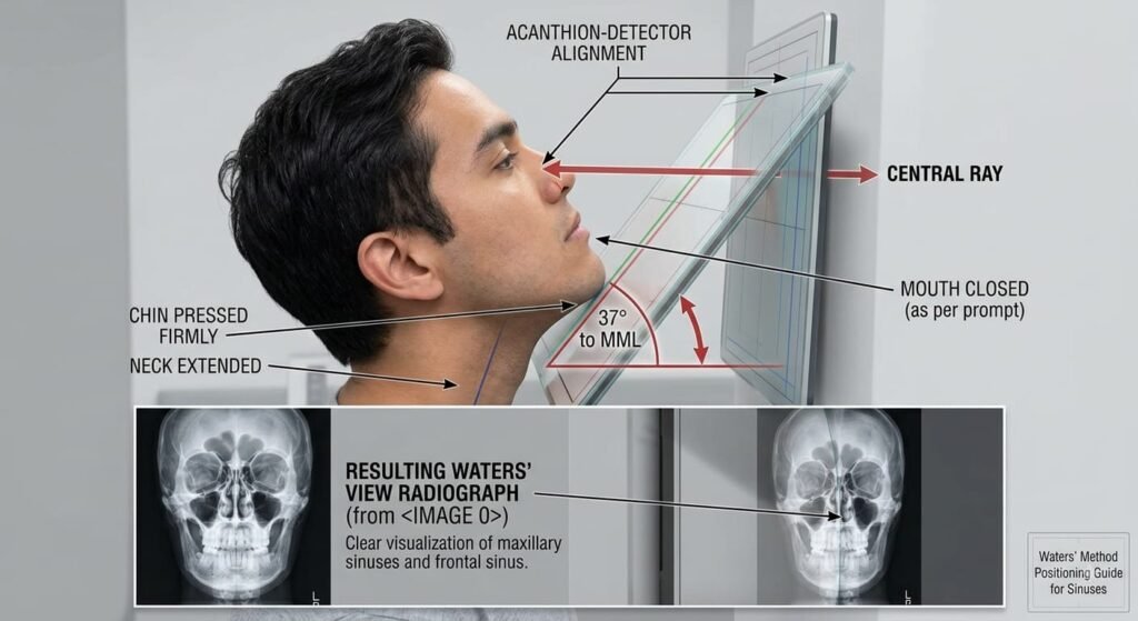
- Standard Waters (Parietoacanthial — Closed Mouth):
- Primary Target: Maxillary sinuses (Antra of Highmore), infraorbital rims, zygomaticomaxillary complexes, and anterior nasal spine.
- Head Extension: Orbitomeatal Line (OML) forms a 37⁰ angle with the Image Receptor (IR); Mentomeatal Line (MML) is perpendicular.
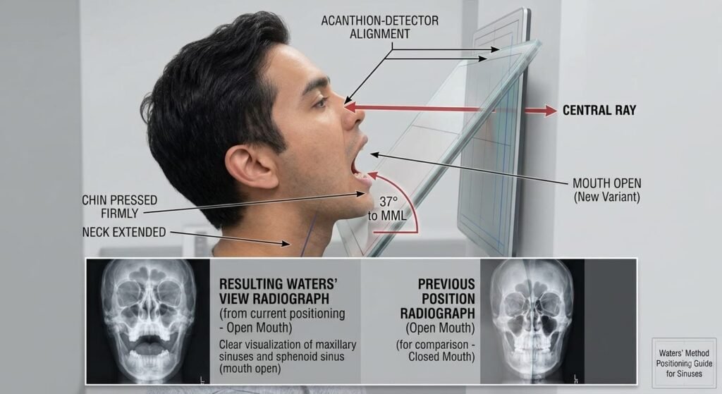
- Transoral Waters (Open-Mouth Waters):
- Primary Target: Sphenoid sinuses projected directly through the open oral cavity without superimposition of the teeth or hard palate, alongside maxillary sinuses.
- Head Extension: Identical 37⁰ OML angle as the standard view, with the mandible dropped maximally.
- Modified Waters (Parietoacanthial — Shallow Angle):
- Primary Target: Orbital floors (detection of blowout fractures and herniated soft tissue) and inferior orbital rims.
- Head Extension: Less neck extension; OML forms a 55⁰ angle with the IR; Lips-Meatal Line (LML) is perpendicular. This places the orbital floor parallel to the Central Ray.
2. Comprehensive Clinical Indications & Pathological Findings
Acute Sinusitis & Air-Fluid Levels
Optimal view for identifying mucosal thickening, polyps, and horizontal air-fluid interfaces within the maxillary antra.
Blowout Fractures (Orbital Floor)
Evaluates orbital floor disruption with herniation of orbital fat or inferior rectus muscle into the maxillary sinus roof.
Tripod / ZMC Fractures
Assesses classic 3-point zygomaticomaxillary fractures: zygomatic arch, infraorbital rim, and lateral orbital margin.
Midfacial Trauma & Le Fort Fractures
Demonstrates lateral nasal wall displacement, nasal bone deviation, and maxillary alveolar separation in facial injuries.
A. Paranasal Sinus Pathology
- Acute Sinusitis:
- Air-Fluid Levels: Sharp, straight horizontal line separating radiolucent air above from radiodense exudate (pus/serous fluid) below. Demonstrable only when the patient is upright with a horizontal beam.
- Mucosal Thickening: Peripheral soft-tissue thickening > 4 mm running along the bony cortical sinus walls.
- Complete Opacification: Total loss of aeration in the maxillary antrum (hazy, white appearance).
- Chronic Sinusitis & Polyposis: Soft-tissue polypoid masses (mucoceles, retention cysts, or sinonasal polyps) showing rounded, convex soft-tissue domes protruding into the sinus cavity.
- Fungal Sinusitis & Mycetoma: Dense, irregular calcifications (antroliths) within an opacified sinus.
- Sphenoid Sinusitis (Open-Mouth): Opacification or fluid levels inside the sphenoid sinuses, critical because untreated sphenoid sinusitis can lead to cavernous sinus thrombosis or intracranial abscess.
B. Maxillofacial & Orbital Trauma
- Blowout Fractures of the Orbit:
- Direct blunt force to the eyeball increases intraorbital pressure, fracturing the paper-thin orbital floor (maxillary sinus roof).
- “Teardrop Sign”: A soft-tissue polypoid density herniating down through the fractured orbital floor into the superior aspect of the maxillary sinus (composed of orbital fat and the inferior rectus muscle, leading to diplopia and enophthalmos).
- Tripod (Zygomaticomaxillary Complex – ZMC) Fractures: Tripartite disruption involving:
- Zygomaticofrontal suture (lateral orbital wall).
- Infraorbital rim and maxillary sinus wall.
- Zygomatic arch.
- Le Fort Midfacial Fractures:
- Le Fort I: Horizontal fracture across the alveolar process of the maxilla.
- Le Fort II (Pyramidal): Extends through the nasal bones, lacrimal bones, infraorbital rim, and maxillary sinus walls.
- Le Fort III (Craniofacial Dysjunction): Complete separation of the facial skeleton from the cranial base traversing the zygomatic arches, orbits, and nasofrontal suture.
- Basilar Skull Fractures (Sphenoid Hemosinus): On Open-Mouth Waters, blood pooling in the sphenoid sinus (hemosinus) serves as an indirect sign of a fracture through the sella turcica or sphenoid bone.
3. Absolute & Relative Contraindications
- Suspected / Uncleared Cervical Spine Trauma:
- Contraindication: Hyperextending the neck to achieve MML perpendicularity can compress or transect the spinal cord if an unstable C-spine fracture (e.g., Jefferson, Hangman’s, or odontoid fracture) is present.
- Trauma Adaptation (Reverse Waters / AP Axial): Keep the patient strictly supine with the head and neck neutral in a C-collar. Angle the Central Ray 30⁰ to 37⁰ Cephalad directed to the acanthion.
- Inability to Sit or Stand Erect (Erect vs. Recumbent Conflict):
- If a patient is placed recumbent (prone/supine) for sinus evaluation, fluid layers along the posterior sinus wall rather than creating a meniscus, causing pseudo-diffuse opacification and masking air-fluid levels.
- Solution: If the patient cannot sit, use a Cross-Table Lateral (Horizontal Beam) to evaluate fluid levels.
4. Technical Factors & Exposure Physics
| Technical Parameter | Standard Waters (Closed/Open) | Modified Waters (Orbits) | Biophysical / Radiographic Rationale |
| Patient Stance | Strictly Erect | Strictly Erect | Allows fluid to settle by gravity; prevents false-negative fluid levels |
| Image Receptor (IR) | 18 × 24 cm (Portrait) | 18 × 24 cm (Portrait) | Tight coverage from frontal sinuses to lower mandible |
| SID | 100 to 115 cm ( 40 to 44 inches) | 100 to 115 cm ( 40 to 44 inches) | Minimizes penumbra and geometric magnification |
| Grid Specification | 8:1 to 12:1focused grid | 8:1 to12:1focused grid | Strips high-angle Compton scatter from dense bone structures |
| Kilovoltage Peak | 70 – 75 kVp | 65 – 75 kVp | High-contrast resolution (k-edge optimized for bone/air borders) |
| Milliampere-seconds | 20 – 30 mAs | 18 – 25 mAs | Calibrated based on facial bone density; center AEC cell active |
| Focal Spot Size | Small (0.6 mm) | Small (0.6 mm) | High spatial resolution for fine orbital lamina papyracea |
| Radiation Shielding | Thyroid & gonadal wrap | Thyroid & gonadal wrap | Protection against primary beam scatter |
| Breathing Phase | Suspended respiration | Suspended respiration | Eliminates respiratory motion and vocal fold movement |
5. Biomechanical Patient Positioning Protocols
Method A: Standard Waters (Parietoacanthial — Closed Mouth)
- Initial Alignment: Seat the patient erect facing the vertical Bucky. Align the midsagittal plane (MSP) perpendicular to the central vertical axis of the grid.
- Head Extension: Have the patient extend their neck, resting the tip of the chin (mentum) firmly against the face of the Bucky.
- Reference Line Check:
- Adjust extension until the Mentomeatal Line (MML) is strictly perpendicular to the IR plane.
- This mechanical orientation ensures the Orbitomeatal Line (OML) forms an exact 37⁰ angle with the receptor surface (or 53⁰ relative to the central ray).
- The tip of the nose should rest approximately 1 to 2 cm off the surface of the Bucky board.
- Tilt & Rotation Check: Ensure the Interpupillary Line (IPL) is parallel to the floor (verifying zero lateral head tilt).
Image Receptor centered to acanthion
(Nose approx. 0.75″ / 2 cm off the board)
(Neck hyperextended)
(Perpendicular baseline reference)
(Aligned with vertical midline)
Enters Parietal Region ➔ Exits at Acanthion
OML 37° / MML Perpendicular
Chin rests on the IR with the neck hyperextended until the MML is perpendicular and the OML forms a 37° angle to the board.
Perpendicular CR to Acanthion
Central ray is directed horizontally at 0° entering posterior parietal bone to exit precisely at the acanthion.
Upright Orientation for Fluid Levels
Always performed with the patient upright whenever possible to demonstrate air-fluid levels in the maxillary sinuses.
MSP Perpendicular (Zero Rotation)
Mid-sagittal plane is aligned perpendicular to the IR to ensure bilateral maxillary antra and orbits are completely symmetrical.
Method B: Transoral Waters (Open-Mouth Parietoacanthial)
- Position the patient identically to the Standard Waters position (Chin on Bucky, MML perpendicular, OML at 37⁰, MSP perpendicular).
- Instruct the patient: “Drop your lower jaw open as wide as possible without moving your head or lifting your chin from the board.”
- Technologist Check: Ensure the patient opens their mouth via mandibular depression only (hinging the TMJ) rather than hyperextending the neck further.
- The open oral cavity allows the horizontal X-ray beam to pass unobstructed beneath the maxillary dentition, penetrating directly through the sphenoid sinuses.
Method C: Modified Waters (For Blowout Fractures)
- Seat the patient erect facing the Bucky.
- Extend the neck slightly less than the standard view, placing both the chin and nose in contact with the Bucky face.
- Adjust the head until the Lips-Meatal Line (LML) is perpendicular to the IR. This sets the OML at a 55⁰ angle to the receptor.
- This positions the orbital floor perpendicular to the IR and parallel to the CR, eliminating perspective distortion of the inferior orbital rim and orbital floor.
6. Central Ray (CR), Beam Alignment & Collimation
- Central Ray Trajectory: Strictly perpendicular (0⁰) to the image receptor and parallel to the floor.
- Exit Point: Directed through the parieto-occipital region to exit precisely at the Acanthion (the midline junction of the upper lip and the base of the nasal septum).
- Field Collimation: Collimated tightly on four sides:
- Superiorly: Just above the frontal sinuses (supraorbital ridge level).
- Inferiorly: Lower margin of the mandibular symphysis (chin).
- Laterally: To the skin margins of the zygomatic arches.
7. Errors, Artifacts & Corrective Actions
| Positioning Error | Morphological Appearance on Film | Pathological Impact | Corrective Action |
| Under-Extension (< 37⁰ / Chin too low) | The petrous ridges project upward into the lower half of the maxillary sinuses. | Obscures the maxillary sinus floors, hiding air-fluid levels and antral fractures. | Elevate the chin further until the MML is perpendicular to the IR. |
| Over-Extension (>37⁰ / Chin too high) | Maxillary sinuses are severely foreshortened; petrous ridges project far below the maxillary alveolar ridge. | Distorts the anatomy of the maxillary antrum and obscures the upper sinus roofs. | Tuck the chin slightly down to restore the 37⁰ OML angle. |
| Lateral Head Rotation | Distance between lateral orbital margins and lateral skull wall is unequal bilaterally; nasal septum tilts away from midline. | Simulates facial asymmetry, pseudo-thickening of walls, and fake zygomatic displacements. | Realign the Mid-Sagittal Plane (MSP) perpendicular to the Bucky. |
| Lateral Cranial Tilt | One orbit/maxillary sinus sits higher than the other; petrous ridges appear at asymmetric heights. | Distorts horizontal baseline; alters horizontal air-fluid line alignment. | Realign the Interpupillary Line (IPL) strictly parallel to the floor. |
| Mandible Closure during Open-Mouth View | Sphenoid sinuses are obscured by the upper incisors, hard palate, and tongue shadow. | Total diagnostic failure to evaluate the sphenoid sinuses. | Ensure maximal oral depression without altering cranial tilt. |
8. Radiographic Anatomy & Quality Evaluation Criteria
Petrous Ridge Projection
Petrous pyramids project strictly inferior to the floor of the maxillary sinuses, clearing the antra entirely.
Maxillary Antra & Septum
Bilateral maxillary sinuses visualized with symmetric aeration; bony nasal septum and vomer aligned along midline.
Orbital Rims & ZMC Arches
Inferior orbital rims, infraorbital foramina, and bilateral zygomatic bones demonstrated without cranial superimposition.
Zero Rotation & Tilt
Equal distance from the lateral orbital margins to the outer cranial borders confirms strict midline alignment.
- Petrous Ridge Clearance (Primary Quality Hallmark):
- The superior borders of the petrous pyramids must be projected immediately inferior to the floor of the maxillary sinuses bilaterally.
- Maxillary Sinuses:
- Symmetrical, unobstructed projection of both maxillary antra with clear aeration and sharply defined cortical bone boundaries.
- Transoral Clearance (Open-Mouth Waters):
- The sphenoid sinuses are clearly visualized within the radiolucent window of the open mouth, unobstructed by the alveolar process of the maxilla or the mandibular teeth.
- Midfacial & Orbital Symmetry:
- Symmetrical infraorbital margins, zygomatic processes, and frontal processes of the maxillae.
- Distance between the lateral orbital rim and the outer lateral calvarium is identical on both sides.
- Nasal septum is vertical and aligned along the central axis without rotational distortion.
- Tissue Contrast: High-contrast resolution distinguishing mucosal soft-tissue swelling from aerated sinus cavities, with sharp, unblurred trabecular detail across all facial bones.
