

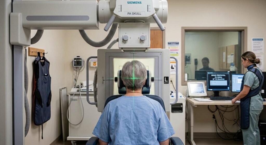
The Posteroanterior (PA) View of the Skull (0° Central Ray) is the fundamental baseline projection for cranial radiography. Unlike the modified Caldwell projection (which uses a 15⁰ caudal tilt), the 0° PA baseline directs the X-ray beam perpendicular to the image receptor, providing an un-distorted view of the frontal bone, internal auditory canals, and symmetrical calvarial structures while minimizing radiation exposure to the eyes.
1. Clinical Indications & Contraindications
Detailed Indications:
- Frontal Bone Pathologies & Trauma: Detection of linear, depressed, or stellate fractures of the frontal bone, supraorbital margins, and frontal air sinuses.
- Calvarial Symmetry & Suture Analysis: Evaluation of the sagittal suture, coronal sutures, and craniosynostosis (premature suture fusion) in pediatric/adolescent cases.
- Neoplastic & Destructive Bone Lesions: Assessment of osteolytic or osteoblastic lesions, Multiple Myeloma (“punched-out” lytic lesions), and metastases in the cranial vault.
- Metabolic & Chronic Bone Diseases: Radiographic diagnosis of Paget’s disease (osteitis deformans), fibrous dysplasia, hyperparathyroidism (“salt-and-pepper” appearance), and hyperostosis frontalis interna.
- Intracranial Calcifications & Foreign Bodies: Identification and localization of radiopaque foreign materials, pineal gland calcifications, choroid plexus calcifications, and falx cerebri calcification.
- Evaluation of Crista Galli & Ethmoid Sinuses: Clear visualization of the perpendicular plate of the ethmoid, crista galli, and superior nasal septum.
Contraindications & Limitations:
- Unstable Cervical Spine Trauma: Absolute contraindication for prone or erect positioning. If a C-spine injury is suspected, never turn the patient face down or manipulate the neck; perform an AP 0° Supine / Trauma AP instead.
- Severe Facial / Nasal Trauma: Patients with active epistaxis, crushed facial bones, or open nasal fractures should not have their forehead and nose pressed against the bucky surface.
- Uncooperative or Bedridden Patients: Severely kyphotic elderly patients or pediatric patients who cannot hold the forehead-nose position should be positioned AP.
- Inferior Orbital Rim Evaluation (Limitation): Because the central ray is 0⁰, the dense petrous ridges completely superimpose over the orbits, obscuring the inferior orbital rims and orbital floor (a 15⁰ Caldwell or 37⁰ Waters view is needed for orbital floor/sinus assessment).
2. Technical Factors
| Parameter | Recommended Specification | Radiographic Rationale |
| Image Receptor (IR) | 24 × 30 cm (10″ × 12″) | Longitudinal / Portrait orientation |
| Source-to-Image Distance (SID) | 100 to 115 cm (40 to 44 inches) | Maintains geometric resolution and controls beam divergence |
| Grid Ratio | 8:1 to 12:1 focused grid | Essential to absorb high amounts of scattered radiation |
| Tube Potential (kVp) | 75 – 85 kVp | High penetration required to pass through dense occipital and petrous bones |
| mAs Range | 25 – 35 mAs | Calibrated based on cranial thickness; center AEC cell active |
| Focal Spot | Small (0.6 mm) | Maximizes spatial detail of thin suture lines and fracture margins |
| Breathing | Suspended respiration | Eliminates respiratory and voluntary motion blur |
3. Step-by-Step Patient & Part Positioning
Cassette top 1.5–2″ above cranial vertex
(Direct contact maintains stability)
(Perpendicular to cassette plane)
(Aligned with vertical grid midline)
Enters Occiput ➔ Exits at Glabella
Forehead & Nose Contact
Patient rests forehead and nose firmly against the upright Bucky surface to lock the cranial baseline.
OML Perpendicular (90°)
Orbitomeatal line is kept strictly 90° to the IR, projecting petrous pyramids across the lower 2/3 of orbits.
MSP Centering (90°)
Mid-sagittal plane is aligned perpendicular to eliminate rotation and maintain bilateral orbital symmetry.
Exit Point (Glabella)
Central ray enters posterior skull at the inion level and exits anteriorly at the glabella along the horizontal axis.
- Patient Preparation: Remove all radiopaque accessories (hairpins, metallic clips, earrings, wigs, spectacles, dentures, hearing aids, and dental plates).
- General Stance:
- Erect (Preferred): Seated or standing facing the vertical Bucky. This is more comfortable and prevents lateral body rotation.
- Prone (Alternative): Patient lies prone on the radiographic table with arms resting comfortably near the shoulders for stabilization.
- Alignment of Reference Lines & Contact Points:
- Forehead & Nose Contact: Rest the patient’s forehead and tip of the nose firmly against the table or vertical Bucky surface.
- Orbitomeatal Line (OML): Adjust head flexion/extension so the OML is strictly perpendicular to the plane of the image receptor.
- Mid-Sagittal Plane (MSP): Align the MSP perpendicular to the midline of the grid/bucky. Stand directly behind the patient to check that the head is not rotated to the left or right.
- Interpupillary Line (IPL): Ensure the IPL is strictly parallel to the plane of the IR (no lateral head tilt).
4. Central Ray (CR) & Collimation
- CR Angle: 0⁰ (perpendicular to the image receptor and parallel to the OML).
- CR Alignment / Exit Point: Directed horizontally (or vertically if prone) to exit at the Glabella (or mid-nasion level).
- Collimation: Four-sided collimation adjusted to the outer skin margins of the skull vault (vertex superiorly, chin base inferiorly, and lateral outer calvarial borders).
5. Key Differences: 0° PA Baseline vs. 15° PA Caldwell vs. 0° AP
| Feature | 0° PA Baseline Skull | 15° PA Caldwell View | 0° AP Skull View |
| Central Ray Angle | 0° (Perpendicular) | 15° Caudad | 0° (Perpendicular) |
| Petrous Ridge Position | Completely fills the orbits (lower 1/3 to middle) | Projected into the lower 1/3 or below orbits | Completely fills the orbits |
| Primary Region Shown | Frontal bone, calvarium, crista galli | Orbital rims, frontal & anterior ethmoid sinuses | Frontal bone & orbits (magnified) |
| Lens Radiation Dose | Minimal (eyes close to IR, away from tube) | Minimal | High (direct exposure to ocular lenses) |
| Orbital Magnification | None / Minimized (Low OID) | Minimized | Increased (Higher OID) |
6. Radiographic Anatomy & Quality Evaluation Criteria
