Dental X Ray radiography
Dental X ray radiography is an essential part of any dental practice. It allows dentists to gain a detailed understanding of the structure and health of the teeth, as well as helping them diagnose disease or injury. This blog post will explore the use of dental X ray radiography, its advantages, disadvantages and safety measures to ensure maximum protection for patients and staff alike. We’ll also look at how this technology has evolved over time, from traditional manual techniques to digital dental X ray systems. Finally, we’ll discuss the implications for practitioners when considering this technology in their practice.
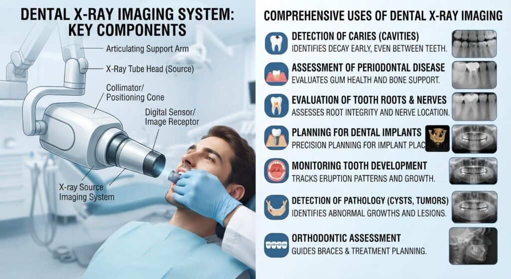
Types
There are two types of dental X-rays: intraoral and extraoral. Intraoral X-rays are taken inside the mouth, while extraoral X-rays are taken outside the mouth. Both types of X-rays are used to diagnose problems with the teeth and jaw.
Intraoral X-rays are the most common type of dental X-ray. They provide a detailed view of the teeth, bones, and soft tissues in the mouth. Intraoral X-rays can be used to diagnose cavities, tooth decay, and other problems with the teeth and jaw. Extraoral X-rays are less common than intraoral X-rays, but they can be used to diagnose problems with the teeth and jaw that cannot be seen with intraoral X-rays. Extraoral X-rays can also be used to assess the status of dental implants and other prosthetic devices.
Uses
There are many benefits to dental X-rays, including the ability to:
-Detect problems early, before they become more serious
-Detect cavities between the teeth that cannot be seen with the naked eye
-Detect hidden tooth decay
-Diagnose periodontal disease
-Diagnose problems with tooth development or alignment
-Detect certain types of tumors
Indication
Dental radiography is used to aid in the diagnosis of dental problems. The most common type of dental radiography is the bitewing radiograph, which is taken by placing the film or sensor in the mouth between the teeth and biting down. This radiograph can show decay between the teeth as well as changes in the bone around the teeth. Other types of dental radiographs include periapical radiographs, which show an image of an entire tooth; panoramic radiographs, which show a wide view of all the teeth; and cephalometric radiographs, which are taken of the side of the head and show all the bones and teeth in profile.
Contraindication
There are certain conditions in which taking dental X-rays is not recommended. These are known as contraindications. Pregnant women, for example, should not have X-rays taken of their teeth because the radiation can harm the developing fetus. People who are allergic to iodine or barium should also avoid X-rays, as these substances are used in the processing of the film.
Dental X ray Tube
Dental X rays are taken using a very small X ray tube. The X ray tube is placed inside the mouth near the teeth that are being examined. The X rays pass through the teeth and are detected by a special film or digital sensor.
Dental X rays are an important part of diagnosing dental problems. They can show cavities, gum disease, infections, and other problems. Dental X rays are typically taken once a year to check for new problems or changes in existing problems.
Dental X ray Procedure
There are two types of dental X-rays: intraoral and extraoral. Intraoral X-rays are taken inside the mouth, while extraoral X-rays are taken outside the mouth. The type of X-ray that is taken will depend on what the dentist is looking for.
Dental X-rays are a vital part of diagnosing and treating dental problems. They allow dentists to see problems that cannot be seen with the naked eye, such as tooth decay, infections, and tumors. Dental X-rays can also help dentists determine the best way to treat a problem.
The procedure for taking a dental X-ray is simple and quick. First, the patient will be asked to bite down on a small piece of film or an electronic sensor. This will help the dentist get a clear image of the teeth and jaws. Next, the X-ray machine will be turned on and aimed at the teeth. The patient will then be asked to hold still while the picture is taken.
Safety
When having a dental X-ray taken, there are a few things you can do to ensure your safety. First, make sure the dental office is using digital X-ray technology. This type of X-ray emits much less radiation than traditional film X-rays. Second, ask the dental team to place a lead apron over your body before the X-ray is taken. This will help protect you from any stray radiation. Finally, be sure to keep your head still during the X-ray so that the image is clear and accurate.
Conclusion
Dental X-Ray radiography is an invaluable tool for dentists, providing them with a detailed view of the inner structures of teeth. It allows for early detection and diagnosis of many dental issues that might otherwise go unnoticed, ensuring better oral health and saving both time and money in the long run. Whereas some patients may find it slightly uncomfortable or invasive, it helps to remember that the benefits far outweigh any potential drawbacks – so make sure you get your regular checkups at your local dentist!