
Caldwell View PNS Radiography: 15° PA Axial Positioning & Anatomy

Caldwell View PNS (15° Caudad PA Axial Projection)
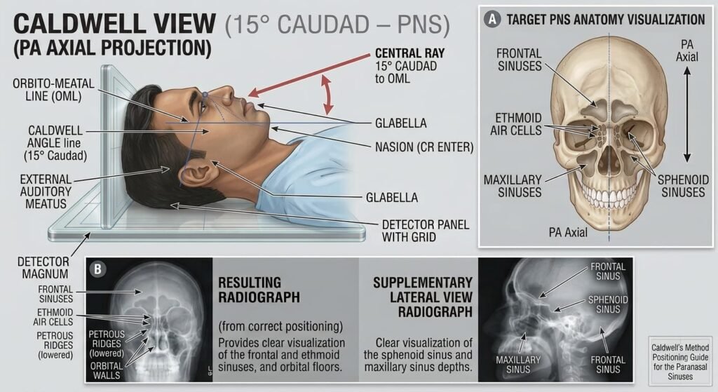
The Caldwell View PNS (15° Caudad PA Axial Projection) is the gold-standard radiographic view designed specifically to evaluate the frontal sinuses and anterior ethmoid air cells. Directing the central ray with a 15⁰ caudal angulation projects the dense petrous ridges of the temporal bones into the lower third of the orbits, leaving the frontal sinuses, orbital margins, and upper two-thirds of the orbits completely unobstructed.
1. Clinical Indications & Contraindications
Detailed Indications:
- Paranasal Sinus Pathologies (Frontal & Ethmoidal):
- Acute & Chronic Sinusitis: Detection of mucosal thickening (>4 mm) along the sinus periphery), air-fluid levels (requires an erect position with a horizontal central beam), and complete sinus opacification.
- Frontal Sinus Mucoceles & Retention Cysts: Expansion, osteolytic thinning, or erosion of the anterior/posterior cortical sinus walls.
- Frontal Sinus Osteomas: Benign, dense radiopaque bone masses arising within the frontal air cavities.
- Maxillofacial & Orbital Trauma:
- Fractures of the superior orbital rim / orbital roof and supraorbital margins.
- Fractures of the lamina papyracea (paper-thin lateral wall of the ethmoid labyrinth).
- Visualization of the superior orbital fissures, foramen rotundum, and the greater and lesser wings of the sphenoid.
- Cranial Symmetry & Bone Lesions:
- Assessment of the crista galli, vertical plate of the ethmoid, and metopic suture closure.
- Congenital hypoplasia or aplasia (absence) of one or both frontal sinuses.
Contraindications & Limitations:
- Unstable Cervical Spine Trauma: Absolute contraindication for prone or forward-flexed erect positioning. If C-spine injury is unconfirmed, perform a Reverse Caldwell View (AP Axial 15⁰ Cephalad) in the supine position without moving the head or neck.
- Maxillary Sinus Floor Assessment (Limitation): The petrous pyramids completely overlap the lower two-thirds of the maxillary antra and nasal fossa. The Waters View is required to evaluate the maxillary sinuses.
- Sphenoid Sinus Evaluation (Limitation): Sphenoid sinuses are superimposed behind the ethmoidal complex and upper nasal cavity.
2. Technical Factors
| Parameter | Recommended Setting | Radiographic Rationale |
| Patient Posture | Strictly Erect | Mandatory for sinus series; gravity is required to demonstrate horizontal air-fluid levels |
| Image Receptor (IR) | 18 × 24 cm or 24 ×30 cm (Portrait) | Tight vertical coverage from frontal sinuses down to the upper jaw |
| SID | 100 to 115 cm ( 40 to 44 inches) | Standard distance to reduce geometric unsharpness and magnification |
| Grid Ratio | 8:1 to 12:1focused grid | Essential to absorb high-angle Compton scatter from cranial bones |
| Tube Potential (kVp) | 70 – 80 kVp | High tissue-to-air contrast resolution for aerated bony sinus walls |
| mAs Range | 20 – 30 mAs | Calibrated based on facial thickness (AEC center cell active) |
| Focal Spot | Small (0.6 mm) | High spatial resolution for fine ethmoid trabeculae and orbital margins |
| Breathing | Suspended respiration | Eliminates respiratory motion blur and vocal cord movement |
3. Step-by-Step Patient & Part Positioning
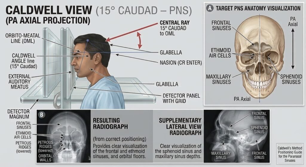
Image Receptor centered to nasion level
(Direct dual-point facial contact)
(Perpendicular baseline alignment)
(Aligned with vertical grid midline)
Enters Occiput ➔ Exits at Nasion
15° Caudad Tube Angulation
Directs the beam angled caudad to project petrous ridges into the lower third of the orbits.
Forehead & Nose Placement
Patient rests forehead and nose firmly against the upright Bucky surface to lock cranial baselines.
OML Perpendicular (90°)
Maintains OML perpendicular to the IR to standardize orbital magnification and petrous ridge descent.
CR Exit at Nasion
Central ray enters posterior occiput and exits directly at the nasion along the sagittal midline.
- Patient Preparation: Remove all metallic and radiopaque objects (hairpins, clips, spectacles, earrings, nose pins, dentures, necklaces, and hearing aids).
- Base Posture: Patient sits or stands erect facing the vertical Bucky with their back straight and shoulders leveled to prevent torso tilting.
- Alignment of Reference Lines & Points:
- Forehead & Nose Contact: Rest the patient’s forehead and the tip of the nose firmly against the surface of the upright Bucky.
- Orbitomeatal Line (OML): Flex or extend the patient’s neck until the OML is strictly perpendicular (90⁰) to the plane of the image receptor.
- Mid-Sagittal Plane (MSP): Must be strictly perpendicular to the midline of the grid (ensures zero left or right head rotation).
- Interpupillary Line (IPL): Must be parallel to the floor and image receptor surface (ensures zero lateral head tilt).
4. Central Ray (CR), Tube Angle & Collimation
- Central Ray (CR) Angle: 15⁰ Caudad (angled downwards toward the feet).
- Alternate Angle Technique (True Calvarium vs. Sinus):
- Standard Caldwell for Sinuses: 15⁰ Caudad (petrous ridges in lower 1/3 of orbits).
- Orbital Fissure / Floor Modified Caldwell: 25⁰ to 30⁰ Caudad (completely clears petrous ridges below the orbital floors).
- CR Entry / Exit Point: Enters the occipital region posteriorly and exits at the Nasion (the midline junction of the frontal and nasal bones).
- Collimation: Four-sided collimation adjusted tightly:
- Superiorly: Just above the upper margin of the frontal sinuses.
- Inferiorly: Below the alveolar ridge of the maxilla.
- Laterally: To the outer skin margins of the orbits and zygomatic bones.
5. Common Positioning Errors & Radiographic Signs
| Error Type | Radiographic Sign on Film | How to Correct |
| Under-Angulation (< 15° or Chin Tucked too much) | Petrous ridges fill the upper half or middle of the orbits, obscuring orbital contents and ethmoids. | Increase the caudal tube angle or check that the OML is strictly perpendicular. |
| Over-Angulation (> 15° or Chin Extended too much) | Petrous ridges project below the inferior orbital rim, causing severe vertical foreshortening of the frontal sinuses. | Decrease the caudal tube angle back to 15⁰ caudad. |
| Head Rotation (MSP not 90°) | Distance from the lateral orbital rim to the lateral skull border is unequal bilaterally; crista galli is displaced from the midline. | Realign the Mid-Sagittal Plane (MSP) perpendicular to the Bucky. |
| Head Tilt (IPL not parallel) | Orbits, frontal sinuses, and petrous ridges are slanted and not on the same horizontal level. | Realign the Interpupillary Line (IPL) strictly parallel to the floor. |
6. Radiographic Anatomy & Quality Evaluation Criteria
Petrous Ridges in Lower 1/3 of Orbits
15° caudad angulation projects petrous pyramids symmetrically into the lower third of the orbital shadows.
Upper 2/3 Orbit & Superior Fissure
Superior two-thirds of the orbits, superior orbital fissures, and supraorbital margins are clearly demonstrated.
Frontal Sinus & Crista Galli
Frontal sinuses are projected unobstructed above the nasal bones; crista galli is centered in the midline.
Midline Cranial Symmetry
Equal distance between the lateral orbital margins and outer cranial borders confirms zero patient rotation.
- Petrous Ridge Projection (Primary Quality Benchmark): Symmetrical petrous pyramids projected cleanly into the lower third of the orbits bilaterally.
- Frontal Sinuses: Completely clear, unobstructed visualization of the frontal sinuses above the nasal bones and crista galli with distinct bony cortical margins.
- Ethmoid Air Cells: Clear visualization of the anterior ethmoid air cells situated between the medial orbital walls and the superior nasal septum.
- Rotational Symmetry:
- Equal distance between the lateral orbital rims and the lateral calvarial borders on both sides.
- Crista galli and the vertical plate of the ethmoid aligned in the midline.
- Tissue Contrast & Sharpness: Sharp trabecular detail across the orbital roofs, supraorbital margins, and ethmoidal labyrinths with no motion artifacts.
