What Is A CT Scan?
Through computer processing, cross-sectional images (slices) of the bones, blood arteries, and soft tissues inside your body are produced during a computerised tomography (CT) scan, which combines a number of X-ray images collected from various angles all over your body. Images from a CT scan offer more information than an X-ray would.
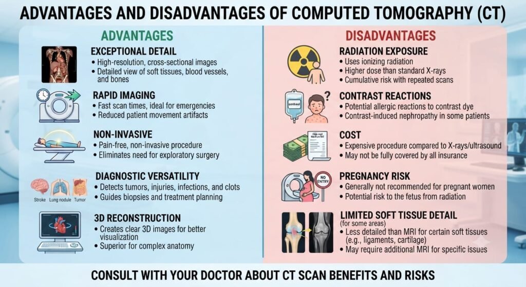
A CT Scan or Computed Tomography scan is an imaging test used to create detailed pictures of the internal organs, bones, and soft tissues inside the body. It can be used to detect anatomical abnormalities and help diagnose various medical conditions and diseases.
How Does A CT Scan Work?
A CT scanner employs a motorised x-ray source that spins around the circular opening of a donut-shaped frame called a gantry, in contrast to a traditional x-ray, which uses a stationary x-ray tube. In a CT scan, the patient is lying on a bed that gently rotates across the gantry as a narrow beam of x-rays is shot into their body by the x-ray tube. Special digital x-ray detectors, which are placed immediately across from the x-ray source, are used in CT scanners in place of film. The detectors catch the x-rays as they leave the patient and send them to a computer.
The CT computer constructs a two-dimensional imaging slice of the patient using advanced mathematical methods each time the x-ray source completes one full revolution. Depending on the CT equipment being used, the thickness of the tissue shown in each imaging slice might change, but it typically ranges from 1 to 10 millimetres . After finishing a full slice, the image is saved and the motorised bed is slowly lowered onto the gantry. Then another image slice is created by repeating the x-ray scanning procedure. Until the necessary number of slices is gathered, this process continues.
The computer can either display the image slices separately or stack them to create a 3D image of the patient that displays the skeleton, organs, tissues, and any anomalies the doctor is hoping to spot. This approach has various benefits, including the ability to rotate the 3D image in space or to see slices one after the other, which makes it simpler to pinpoint the precise location of a potential problem.
Why Is a CT Scan Carried Out?
A CT scan can provide a more accurate and detailed view of the body’s internal structures than a standard X-ray. This makes it an invaluable tool for diagnosing a wide range of conditions, such as:
- Cancer
- Diagnose muscle and bone disorders, such as bone tumors and fractures
- Internal Bleeding
- Pinpoint the location of a tumor, infection or blood clot
- Heart Disease
- Guide procedures such as surgery, biopsy and radiation therapy
- Brain Aneurysms
- Detect and monitor diseases and conditions such as cancer, heart disease, lung nodules and liver masses
- Infections
- Monitor the effectiveness of certain treatments, such as cancer treatment
- Abdominal Masses
- Detect internal injuries and internal bleeding
- Pulmonary Embolism
- Bone Injuries
What Are The Risks of CT Scan?
A CT scan normally carries very low risks. The scan itself is non-invasive and painless, and the radiation dose is extremely low and poses no harm to the patient. However, in some cases, a contrast material may be injected into a vein to obtain better imaging results. This can cause side effects, such as nausea and allergic reactions, in some patients. If you are pregnant, let your doctor know. If you require imaging of the stomach region, your doctor could advise an ultrasound or another non-ionizing scan.
X-rays, which create ionising radiation, are used in CT scans. According to research, this type of radiation may harm your DNA and cause cancer. However, the possibility of getting a deadly malignancy as a result of a CT scan is still extremely low—roughly it’s 1 in 2,000.
CT scans can have some risks, though serious side effects are rare when the procedure is performed correctly. Possible risks of CT scans can include an increased risk of cancer due to radiation exposure, an allergic reaction to contrast material, or a complication from intravenous contrast material, such as kidney damage.
However, the effects of radiation accumulate over time. As a result, each CT scan you get raises your risk. Ask your doctor why the CT scan is required and discuss any potential risks and advantages of the procedure.
In youngsters, ionising radiation can be more damaging. This is a result of their ongoing growth. Additionally, they will have more time to be exposed to radiation. Before the treatment, you might wish to find out if the settings on the CT scanner have been changed to accommodate children.
Overall, CT scans offer a fast, accurate way to diagnose medical conditions and can help doctors decide on the best course of treatment for their patients.
What is a CT scan used for?
CT scans are used to diagnose a variety of medical conditions and to examine the body for changes. They are used to detect tumors, cysts, injury, infection, inflammation and many other diseases and general medical conditions. They are also used to check for abnormal structures and monitor the progress of certain medical conditions, such as cancer. Additionally, a CT scan is often used to guide certain medical procedures, such as biopsies.