IVU (Intravenous urogram) or IVP (Intravenous pyelogram)-
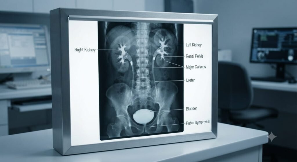
After a contrast dye has been injected into a vein, an x-ray picture of the prostate, kidneys, ureters, and bladder is obtained. These organs become dazzling white when the dye accumulates in them throughout its journey via the circulation. This enables the medical professional to spot obstructions or other urinary tract issues on the x-ray images. Certain urinary system diseases, such as kidney or bladder stones, kidney cysts, urinary tract tumors, and an enlarged prostate, may be diagnosed with the aid of an IVU. Also known as IVP, intravenous pyelogram, and intravenous urogram.
An intravenous urogram (IVU) is a test that looks at the whole of your urinary system. It’s sometimes called an intravenous pyelogram (IVP). It looks at the:
- kidneys
- bladder
- tubes that connect the kidney with the bladder (ureters)
Terminology –
Others reserve the name “pyelogram” for retrograde investigations involving the collecting system and prefer to use the term “urogram” to describe the visualisation of the kidney parenchyma, calyces, and pelvis following intravenous injection of contrast. Both phrases are frequently interchanged in real-world situations.
Indication-
- check for normal function of kidneys
- flank and lower back pain and hematuria
- congenital anomalies of the urinary tract
- check the course of the ureters
- urinary calculi
- enlarged prostate
- neoplasms of kidney
- strictures of the urinary tract.
- detect and localise a ureteric obstruction (urolithiasis)
Contraindication-
- You have an iodine or contrast dye allergy.
- Your kidneys are sick.
- You may be pregnant or you are. Given that X-rays only expose the patient to a brief radiation burst, your doctor will probably choose for a different test.
- Children and infants rarely get IVPs.
Patient Prepration-
- 5 hours of overnight fasting before the examination; Given that laxatives do not enhance image quality, they may not be essential for bowel preparation.
- Take a scout/pilot film the day of the surgery to assess the patient’s preparation and look for radiopaque calculi.
- The calcification fields must be assessed before to the infusion of contrast. If not, the contrast can mask the calcification, which could lead to missed results.
- Radiographs taken at an angle can assist confirm the location and kind of calcifications. When the patient may have urinary tract calculus symptoms but none are otherwise visible on the AP view, this view is crucial.
- Verify if the serum creatinine level is within the normal range (as per hospital guidelines)
- Inform your doctor in advance of the test if you are expecting, suffer from any ailments, have allergies, or use any prescription or over-the-counter medications.
- Before your procedure, you might need to stop taking any of the following:
- Blood thinners like aspirin and diabetic medication like metformin
- There should always be emergency supplies and medicines on hand in case a patient reacts to contrast.
Technique-
Exposures typically range from 65 to 75 kV, 600 to 1000 mA, for <0.1 second. The renal parenchyma exhibits less contrast at higher kV levels.
Procedure-
The following phases make up the conventional pyelography process.
- Next, a bolus of contrast should be administered.
- For the administration of a water-soluble contrast, IV access is necessary. The desired contrast is nonionic.
- The dose will vary depending on the patient’s weight; typically, the patient can tolerate up to 1.5 ml/kg of body weight.
- The contrast dosage is typically injected quickly (bolus)
- 10 to 14 seconds after contrast administration, an instantaneous AP film is obtained to see the renal parenchyma/outline for masses.
- Nephrographic pictures should be taken 1 to 3 minutes after contrast delivery. (Oblique image are an additional choice.)
- After administering contrast for five minutes, a KUB radiograph should be taken.
- After receiving a KUB radiograph, immediately begin abdominal compression.
- For the purpose of seeing the ureters and pelvicalyceal systems as needed and at the operator’s discretion, serial radiographs are acquired every 5 to 20 minutes.
- Obtain a KUB radiograph and fluoroscopic spot pictures of the ureters right away after the compression has been released.
- Obtain the bladder’s radiographic picture.
Compression is contraindicated in-
renal traumalarge
abdominal mass
abdominal surgery (post operative)
abdominal aortic aneurysm
Potential Diagnosis-
Assessment of both kidneys
- Size
- Cyst
- Position
- Renal parenchyma and parenchymal thickening
- Abortive calyces
- Rounding of forniceal margins
- Vertical axis
- Aberrant papillae
- Renal contour and symmetry
- Scarring of the parenchyma
- Calyceal distortion
- Double contouring of renal parenchyma
- Loss of papillary impression
Assessment of the ureters
- Symmetry of both the ureter(s)
- Ureteral obstruction
- Diverticulum/diverticula of ureter(s)
- Medial and lateral deviation of the ureter(s)
- Obstructive and non-obstructive dilatation of ureter(s)
- Asymmetry of the ureteral caliber
Assessment of the urinary bladder
- Physiologic distention
- Contour abnormalities
- Diverticula
- Bladder wall thickening
- Neoplasms
- Irregularity of the lumen
- Position of the bladder
Complications-
During Preparation-
- The patient is instructed to fast before surgery.
- The patient must empty their bladder before to surgery.
- Mild laxatives may be prescribed by the doctor.
- To the sufferer: Explain the process.
- Take note of the patient’s comorbidities, allergies, prior health issues, and drug history.
- Ask the patient to remove all jewellery and other metal accessories prior to the procedure.
- Make that the patient, if a woman, is not pregnant when the procedure is performed.
- Take precautionary measures to shield the unborn child from radiation exposure if she is pregnant.
Usually, this test is done without hospitalisation.
- The patient must stay immobile on the treatment table the whole time.
- The procedure usually takes an hour, but if the kidneys are not functioning properly, it might take longer.
- To properly visualise the lower urinary tract segments, compression bands could be required.